


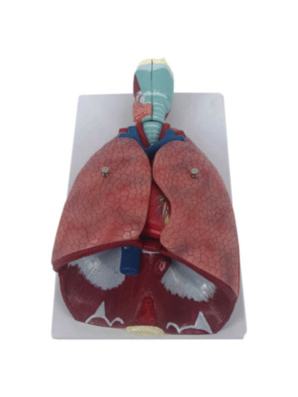
ATL-196 HUMAN THORACIC ORGANS (ANAT-MAGNIFIED MODEL OF LARYNX HEART AND LUNGS )SOFT
The lung model with larynx is first class. The high quality lung model contains the following removable parts for added anatomical detail: * 2-part larynx * Trachea with bronchial tree * 2-part heart * Subclavian artery and vein * Vena cava * Aorta * Pulmonary artery * Esophagus * 2-part lung (front halves removable) * Diaphragm. This is a great model of the anatomy of the lung area.
Classification:
Respirotary System (nose, throat, lung, trachea, etc.)
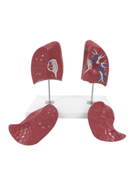
ATL-197 HUMAN LUNGS 2 PARTS
This lung model clearly represents the lung structures of both the left and right lobes; showing both characteristics and differences. Not only is the lung model suitable for explaining the anatomy of the lungs within the contexts of a biology class, but it is also great for explaining anatomical details, diseases etc. to patients.
Classification:
Respirotary System (nose, throat, lung, trachea, etc.)
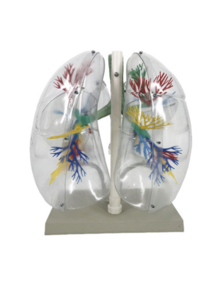
ATL-198 MODEL OF TRANSPARTENT HUMAN LUNG SEGMENT (JUMBO)
This lung model clearly shows the structure of a set of lungs, thanks to its transparent outer construction. This transparent lung model has been especially designed to show the structure of the lungs in order to facilitate visualisation of their anatomy, both for teaching and demonstration. For this purpose, the external anatomy of the lung model is composed of transparent plastic and both the lungs and bronchial tree are composed of segments–each of a different colour.
Classification:
Respirotary System (nose, throat, lung, trachea, etc.)
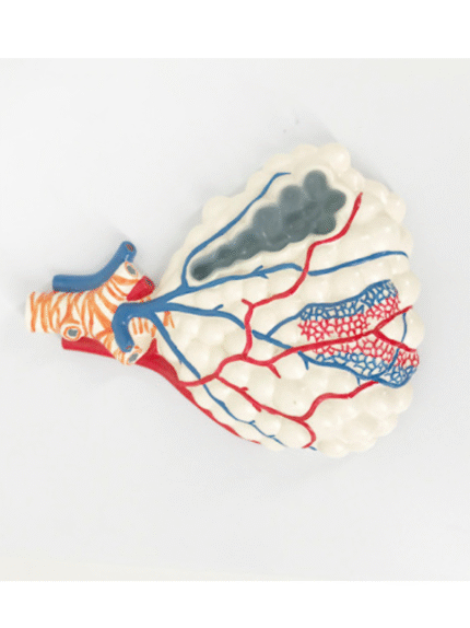

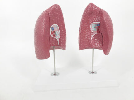
ATL-199 MAGNIFIED PULMONARY ALVEOLI MODEL
The model shows the branching of bronchioles, respiratory bronchioles, alveolar ducts, alveolar sacs, and alveoli. The alveolar sacs show their cross-sectional structure, and display bronchial arteriovenous, pulmonary arteriovenous, capillary network, pulmonary membrane, smooth muscle, elastic fibers, and reticular fibers.
Classification:
Respirotary System (nose, throat, lung, trachea, etc.)
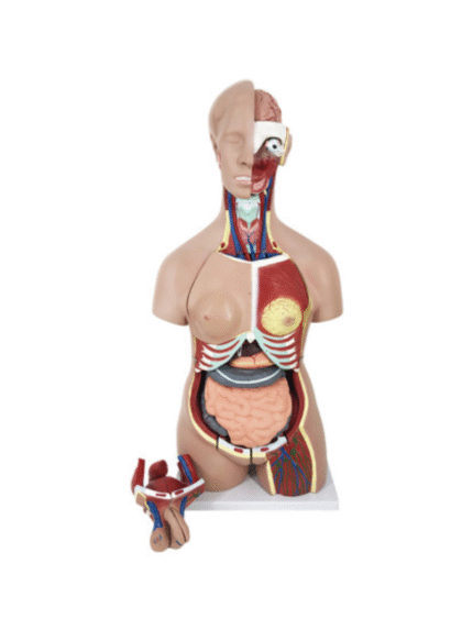
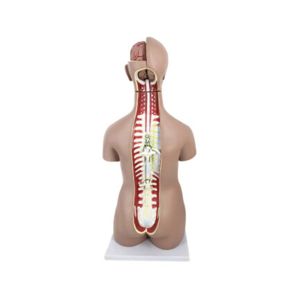
ATL-20 ADVANCED MODEL OF HUMAN TORSO UNI-SEX 45 CM 23 PARTS
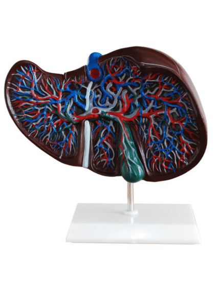
ATL-200 HUMAN LIVER GAINT
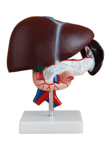
ATL-201 HUMAN LIVER PANCREAS &DUODENUM

ATL-202 PANCREAS WITH SPLEEN AND DUODENUM SOFT
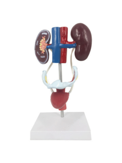
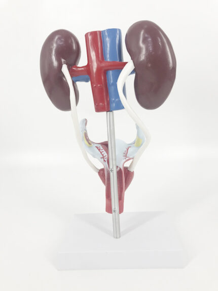
ATL-203 HUMAN FRMALE UROGENTIAL SYSTEM
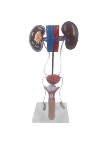
ATL-204 HUMAN MALE UROGENITAL SYATEM
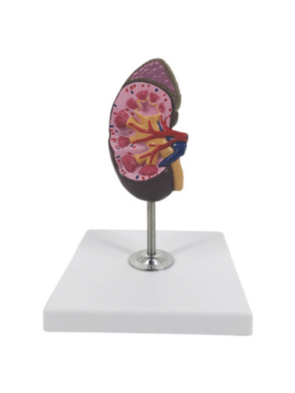
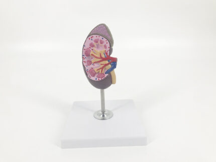
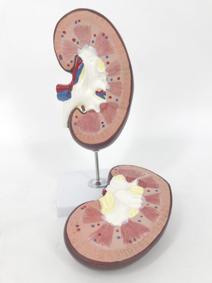
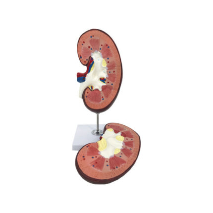
ATL-205 HUMAN KIDNEY 1 PARTS
This model of a human kidney in almost life size shows hand painted details of renal pelvis, renal medulla, renal calyx, renal cortex, renal artery and vein, ureter and adrenal gland.
Classification:
Pathological & Promotional Models
Urinary system (kidney, bladder, pelvic cavity, male and female genitals, etc.)