


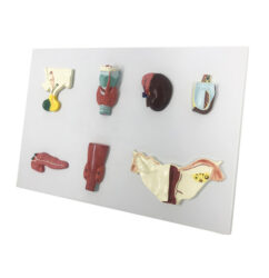
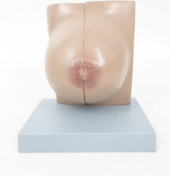


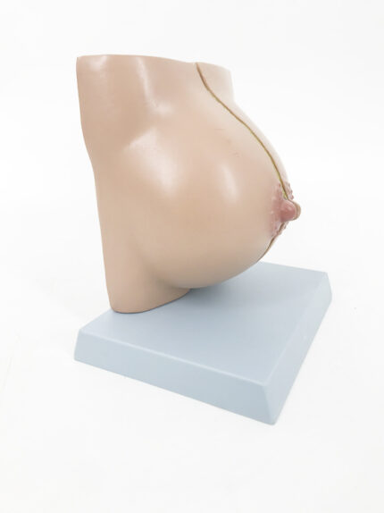
ATL-143 MAMMERY GLAND IN RESTING PERIODS
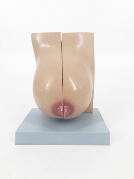
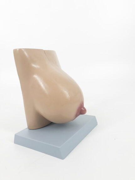
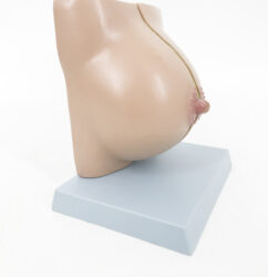
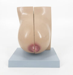
ATL-144 MAMMY GLAND IN LACATION
Classification:
Pathological & Promotional Models
Urinary system (kidney, bladder, pelvic cavity, male and female genitals, etc.)
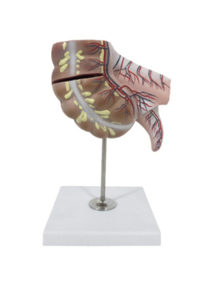
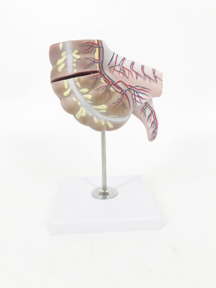
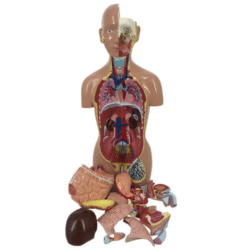
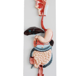
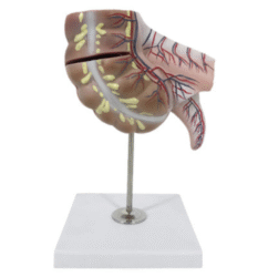
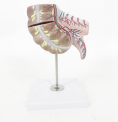
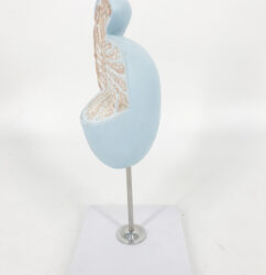
ATL-146 APPENDIX AND CAECUM SOFT
Classification:
Pathological & Promotional Models
Digestive System(oral cavity, stomach, liver, pancreas, intestine, etc.)
ATL-147 MAGNIFIED TESTICLE SOFT
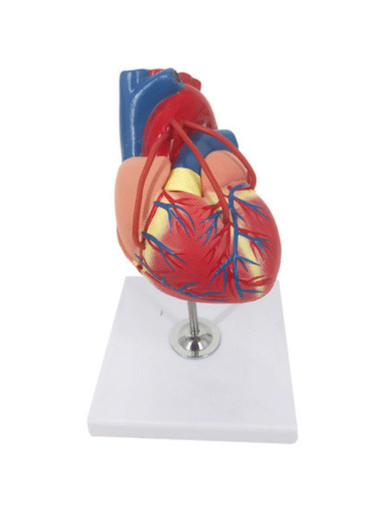

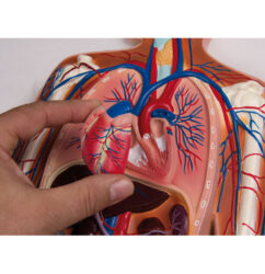
ATL-156 ADVANCED ANATOMICAL MODEL OF HUMAN HEART SOFT
Classification:
Pathological & Promotional Models
Blood Circulation System(heart, blood cells, blood vessels, etc.)
ATL-161 THE DEVELOPMENT MODEL OF HUMAN HEART (SET OF 12 )
Classification:
Pathological & Promotional Models
Blood Circulation System(heart, blood cells, blood vessels, etc.)
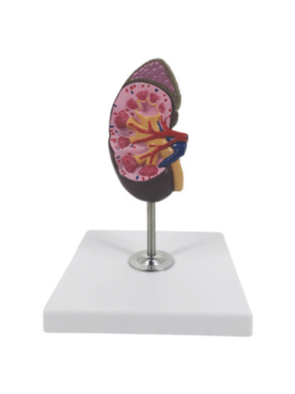
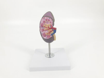
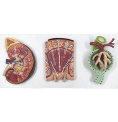
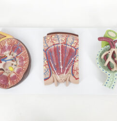
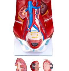
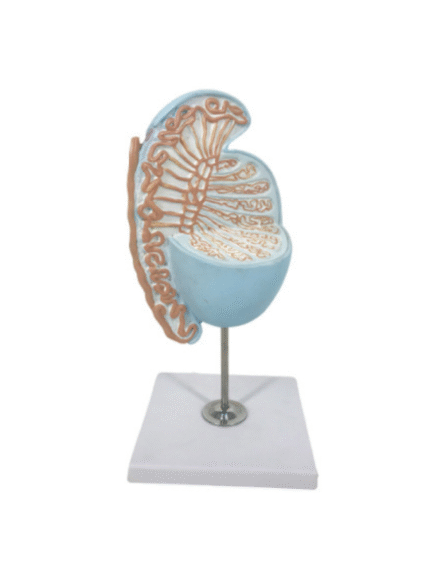
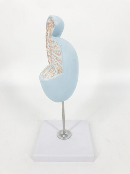
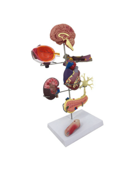
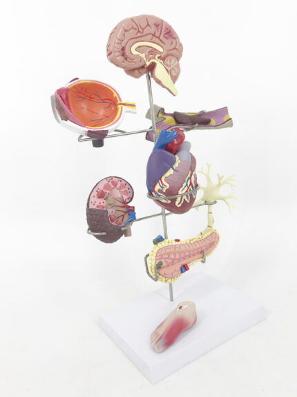
ATL-205 HUMAN KIDNEY 1 PARTS
Classification:
Pathological & Promotional Models
Urinary system (kidney, bladder, pelvic cavity, male and female genitals, etc.)
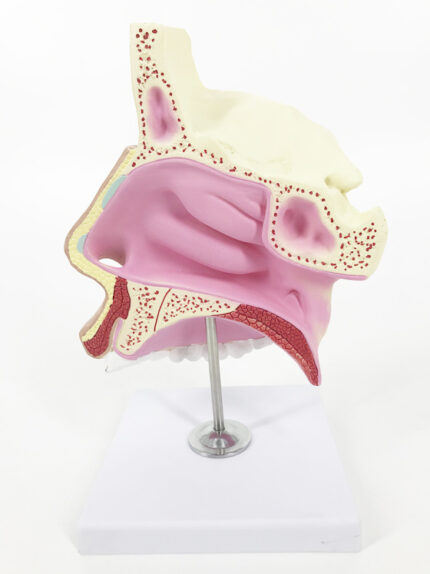

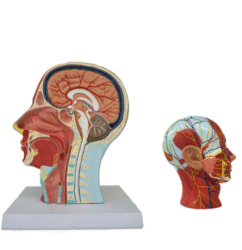
ATL-216 HUMAN NASAL CAVITY
Classification:
Pathological & Promotional Models
Respirotary System (nose, throat, lung, trachea, etc.)

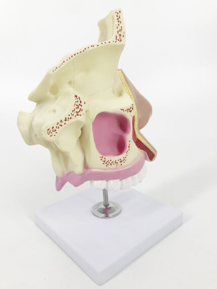
ATL-216 HUMAN NASAL CAVITY
Classification:
Pathological & Promotional Models
Respirotary System (nose, throat, lung, trachea, etc.)