


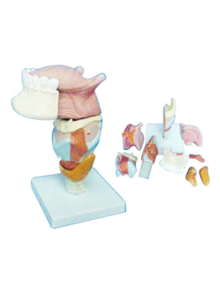
ATL-194 LARYNX WITH TONGUE AND TEETH SOFT
This item dissects into 5 different pieces. To make it easier to pull each piece apart, the product includes magnets that are strong enough to keep the model together on display, but easy to remove. Beginning with the base, the model extends from the trachea to the tongue. This model also provides a detailed look at sublingual gland and sub-mandibular gland as well as the vocal cord and epiglottis.
Classification:
Respirotary System (nose, throat, lung, trachea, etc.)


ATL-195 HUMAN LARYNEX
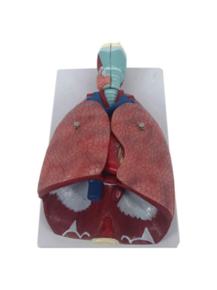
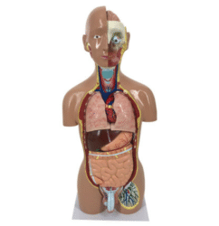
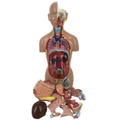
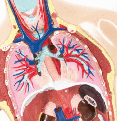
ATL-196 HUMAN THORACIC ORGANS (ANAT-MAGNIFIED MODEL OF LARYNX HEART AND LUNGS )SOFT
The lung model with larynx is first class. The high quality lung model contains the following removable parts for added anatomical detail: * 2-part larynx * Trachea with bronchial tree * 2-part heart * Subclavian artery and vein * Vena cava * Aorta * Pulmonary artery * Esophagus * 2-part lung (front halves removable) * Diaphragm. This is a great model of the anatomy of the lung area.
Classification:
Respirotary System (nose, throat, lung, trachea, etc.)
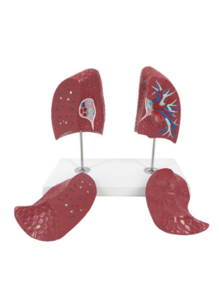
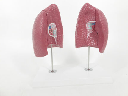
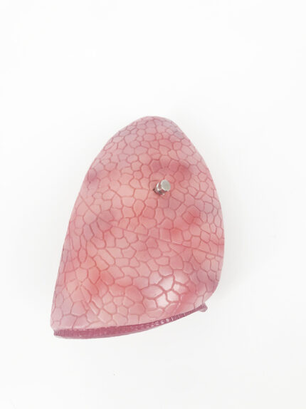
ATL-197 HUMAN LUNGS 2 PARTS
This lung model clearly represents the lung structures of both the left and right lobes; showing both characteristics and differences. Not only is the lung model suitable for explaining the anatomy of the lungs within the contexts of a biology class, but it is also great for explaining anatomical details, diseases etc. to patients.
Classification:
Respirotary System (nose, throat, lung, trachea, etc.)
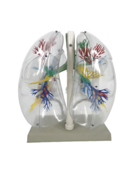
ATL-198 MODEL OF TRANSPARTENT HUMAN LUNG SEGMENT (JUMBO)
This lung model clearly shows the structure of a set of lungs, thanks to its transparent outer construction. This transparent lung model has been especially designed to show the structure of the lungs in order to facilitate visualisation of their anatomy, both for teaching and demonstration. For this purpose, the external anatomy of the lung model is composed of transparent plastic and both the lungs and bronchial tree are composed of segments–each of a different colour.
Classification:
Respirotary System (nose, throat, lung, trachea, etc.)
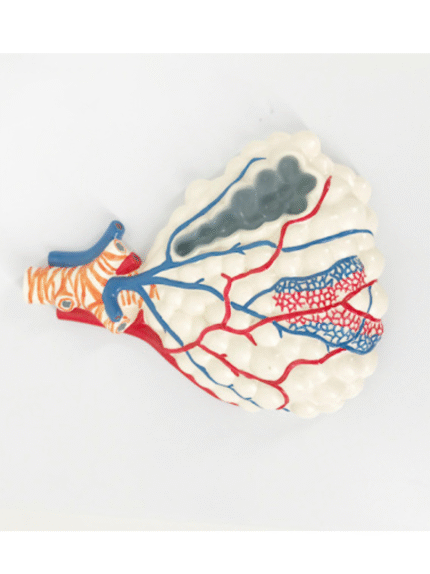

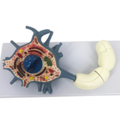
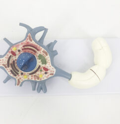
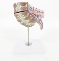
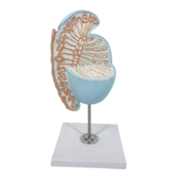
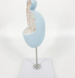
ATL-199 MAGNIFIED PULMONARY ALVEOLI MODEL
The model shows the branching of bronchioles, respiratory bronchioles, alveolar ducts, alveolar sacs, and alveoli. The alveolar sacs show their cross-sectional structure, and display bronchial arteriovenous, pulmonary arteriovenous, capillary network, pulmonary membrane, smooth muscle, elastic fibers, and reticular fibers.
Classification:
Respirotary System (nose, throat, lung, trachea, etc.)
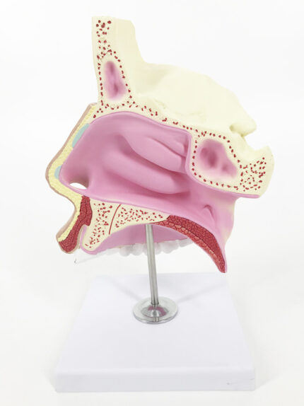

ATL-216 HUMAN NASAL CAVITY
This life size model shows a section of the nasal cavity:1. External nose: showing the nasal bone and nasal cartilage ofthe cut.2. nasal cavity: the lateral wall of the upper, middle and lowerthree turbinate into the nasal cavity, the formation of the upper,middle and lower three nasal passages.3. nasal sinus: frontal sinus, sphenoid sinus and maxillary sinus.
Classification:
Pathological & Promotional Models
Respirotary System (nose, throat, lung, trachea, etc.)

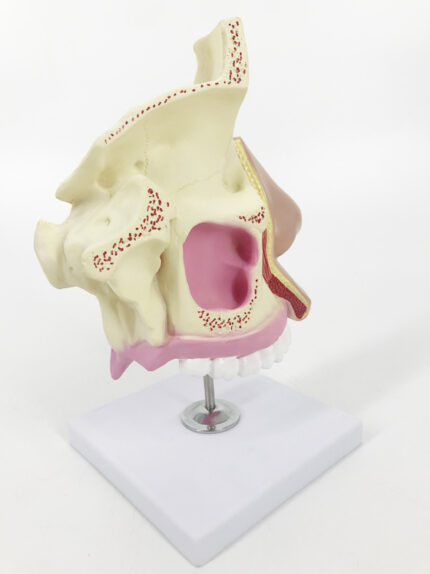
ATL-216 HUMAN NASAL CAVITY
This life size model shows a section of the nasal cavity:1. External nose: showing the nasal bone and nasal cartilage ofthe cut.2. nasal cavity: the lateral wall of the upper, middle and lowerthree turbinate into the nasal cavity, the formation of the upper,middle and lower three nasal passages.3. nasal sinus: frontal sinus, sphenoid sinus and maxillary sinus.
Classification:
Pathological & Promotional Models
Respirotary System (nose, throat, lung, trachea, etc.)