


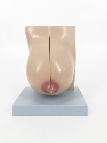
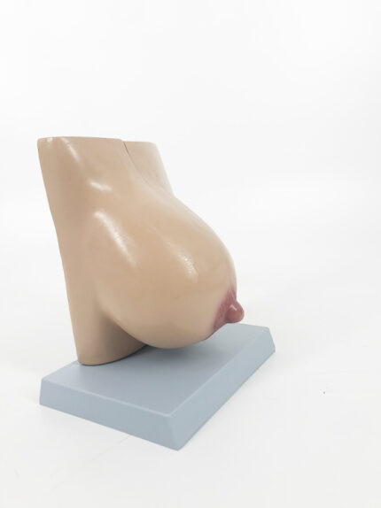
ATL-144 MAMMY GLAND IN LACATION
This model opens in half to display all internal structures of a female breast during lactation. It is able to stay closed by a metal hook, and is mounted on a secure white base for easy display and storage.
Classification:
Pathological & Promotional Models
Urinary system (kidney, bladder, pelvic cavity, male and female genitals, etc.)
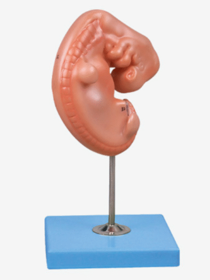
ATL-145 EMBRYO SOFT
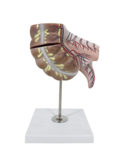
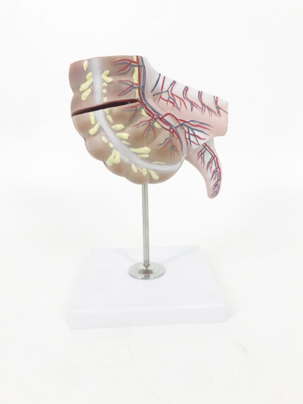
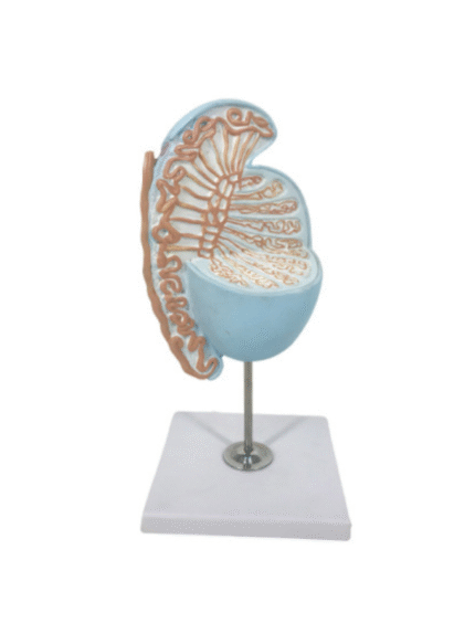
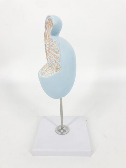
ATL-146 APPENDIX AND CAECUM SOFT
This anatomical model is painted in a natural color tone, with extensive detailing of the caecum, which is generally considered the beginning of the large intestine. Markings include walls of the intestine as well as veins and arteries which deliver blood flow to the large intestine. Below the caecum, the appendix extends from the model, demonstrating the location and attachment of the appendix to the large intestine. This anatomy model can be opened to explore greater, internal detail of the large intestine, with accurate depiction of the folds and texture of the internal anatomy of the intestine. The removable portion of the model is secured with magnets, allowing for a clean, uninterrupted view of the intestinal anatomy.
Classification:
Pathological & Promotional Models
Digestive System(oral cavity, stomach, liver, pancreas, intestine, etc.)
ATL-147 MAGNIFIED TESTICLE SOFT

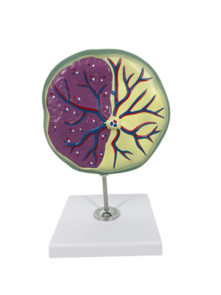
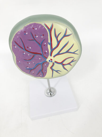
ATL-148 ENLARGED MODEL OF PLACENTA SOFT
This model describes the anatomical structures of the human placenta in a cross-section view. This enlarged model is useful to learn about how the placenta works to provide nutrition, oxygen, blood and waste elimination between a developing fetus and the uterine wall.
Classification:
Hietology, Mbryology Models(skin, embryo, etc.)
ATL-149 PLACENTA MODEL SOFT
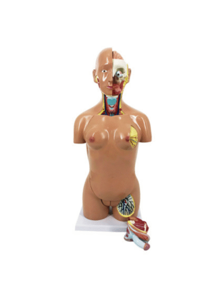
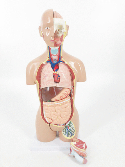
ATL-15 HUMAN TORSO FEMALE 85 CM WITH ORGANS 19 PARTS HARD ORGANS
This life size model with open back is composed of 18 parts andprovides an exceptionally realistic reproduction of anatomical structures in their finest detail. All of the body system arerepresented with fully accessibility.Torso*1 Brain*1 Eyeball*1 Lung*2Heart*2 Liver*1 Stomach*2 Intestine*2Male genital organ*2 Female genital organ*2Breast cover*1 Vertebrea*1
Classification:
Human Torso Models (human torso, fault, etc.)
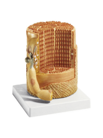
ATL-150 SKELETAL MUSCLE FIBER WITH MOTOR END-PLATE
This micro-anatomy model magnifies the anatomy of the human muscle fiber approximately 10,000 times. This muscle model illustrates a section of a skeletal muscle fiber and its neuromuscular end plate. The muscle fiber is the basic element of the diagonally striped skeletal muscle.
Classification:
Hietology, Mbryology Models(skin, embryo, etc.)

ATL-151 SPINAL CORD IN THE SPINAL CANAL
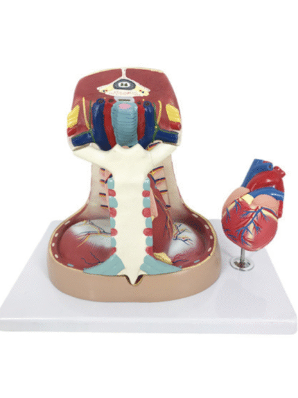
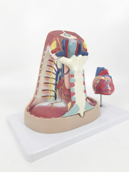
ATL-152 MEDIASTIUM MODEL
This life size model is composed of 5 parts, including a 2-part heart that provides an interior view of the chambers and valves. The sternum and thymus are removable to reveal the pericardical sac and the major pulmonary and systematic vessels. The trachea and esophagus are shown entering the mediastinum through the superior thoracic aperture; the inferior thoracic aperture is delimitated from the diaphragm musculature.
Classification:
Human Trunk Models (head, hand, foot, chest cavity, perineal, etc.)

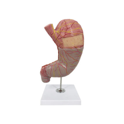
ATL-153 HUMAN STOMACH SOFT
This model is dissected along the medial plane and can be opened to show the internal structure of the stomach, including the mucosa, the pylorus and a section of the gastric wall. Superficial dissections expose the longitudinal, circular and oblique muscle layers, with nerves and vascular structures.
Classification:
Digestive System(oral cavity, stomach, liver, pancreas, intestine, etc.)
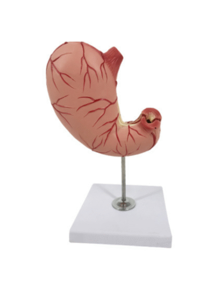
ATL-154 HUMAN STOMACH
This life size model is dissected along the medial plane and can be opened to show the internal structure of the stomach, including the mucosa, the pylorus, a section of the gastric wall. The model also shows the superficial muscular layers and the blood vessels.
Classification:
Digestive System(oral cavity, stomach, liver, pancreas, intestine, etc.)